
NEWS
An MRI Score Times Decline in Early-Onset Alzheimer’s
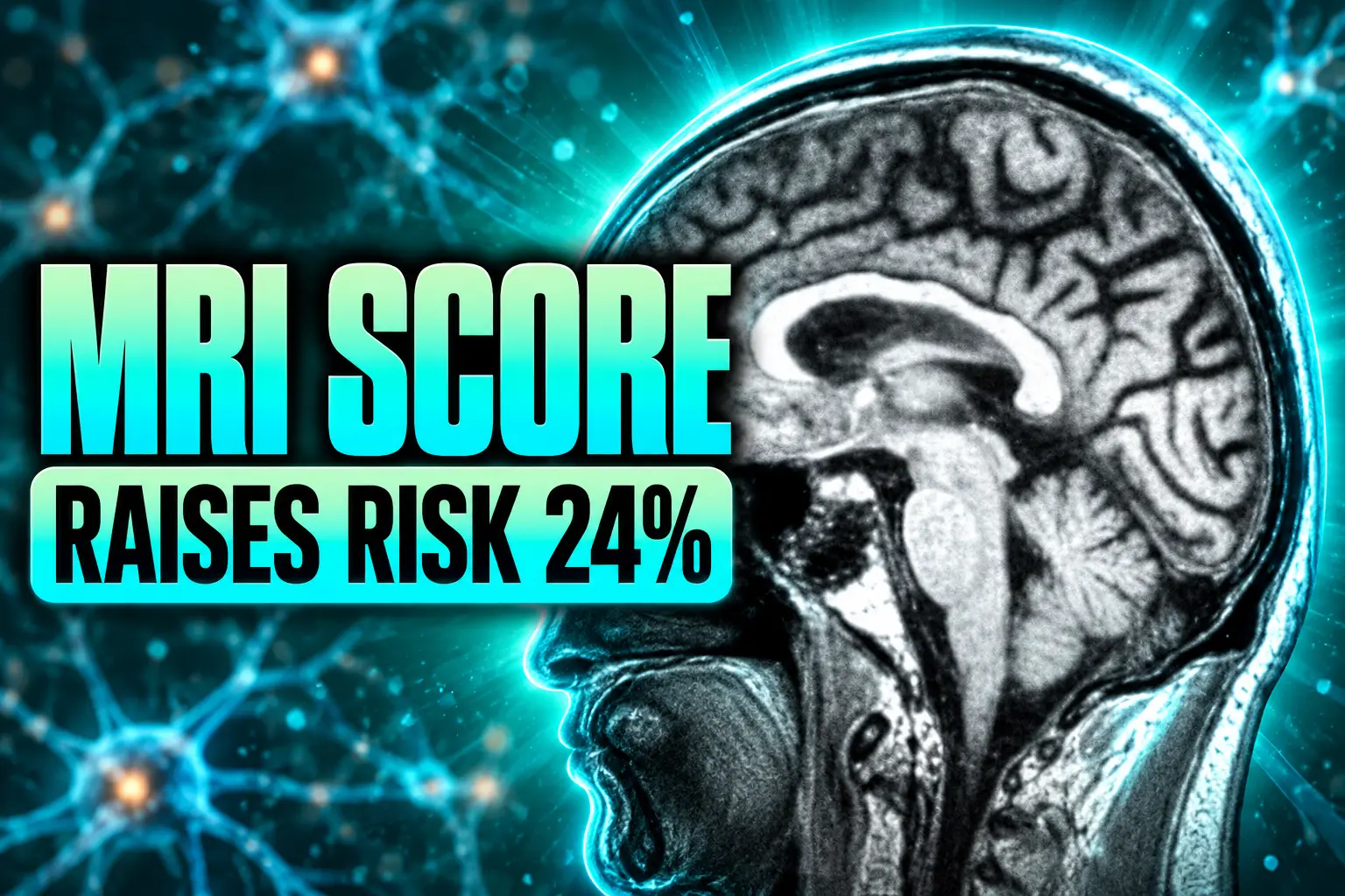
A Neurology study found that shrinkage in eight thinking regions, not the hippocampus, raised dementia risk 24 percent per standard deviation in early-onset.

An MRI shrinkage score raised dementia risk by 24 percent per standard deviation in 130 people with early-onset Alzheimer’s disease. Mass General Brigham researchers published the finding August 26, 2026, in Neurology, using scans already taken in a U.S. study of patients aged 40 to 64 who still lived independently.
The score does not replace amyloid or tau tests. It tries to pull a timeline from a scan most of these patients already receive, which is the piece families have been asking clinics to give them.
Two-Thirds Reached Dementia in About Two Years
Thiago Paranhos, M.D., of Massachusetts General Hospital and Harvard Medical School, and colleagues followed 130 people with biomarker-supported sporadic early-onset Alzheimer’s at the mild cognitive impairment stage, plus 97 cognitively normal controls of similar age. Patients’ mean age at the first scan was 59.6 years, and 49 percent were women. All started at a global Clinical Dementia Rating of 0.5, meaning they had thinking problems but still managed daily life.
Mean follow-up was 21.73 months. In that window, 84 of 130 patients, about 65%, progressed to dementia, recorded as the first visit with a global CDR of 1, when memory, language, or problem solving begins to interfere with independence. The 46 who stayed at MCI were followed a little longer, about 24 months, than those who converted.
| Baseline measure | Progressors (n = 84) | Stayed at MCI (n = 46) |
|---|---|---|
| Age at scan, years | 59 | 60.65 |
| Share of men | 43% | 65% |
| CDR sum of boxes | 3.03 | 2.12 |
| Mini-Mental State Examination | 23.05 | 25.80 |
Progressors were younger, more often women, and already more impaired on both the CDR sum of boxes and the MMSE, the paper reported. Education did not differ. Amyloid PET centiloid values in the patient group averaged 97.17, against 6.17 in controls, so the sample was not a mixed bag of look-alike dementias.
On the baseline MRI, greater gray-matter thinning in a preset set of thinking regions predicted a faster move to dementia. The univariable hazard ratio of 1.24 per standard deviation of extra shrinkage carried a 95 percent confidence interval of 1.13 to 1.37. After age, sex, and baseline CDR-SB were in the model, the MRI term still predicted conversion, with a hazard ratio of 1.14.
Hippocampal Volume Missed the Signal
Memory clinics still lean on hippocampal volume because that is the late-onset playbook. In this early-onset cohort that measure did not predict who lost independence. The hippocampal hazard ratio was 1.21, with a p value of 0.0728, so it stayed on the wrong side of the study’s significance line.
Whole-brain cortical thickness did predict conversion (hazard ratio 1.31), as did several large-scale networks. Among those models, the early-onset signature had the best fit. The right midlateral temporal cortex, one piece of that signature, posted the highest single-region hazard ratio, 1.46.
| MRI measure | Hazard ratio | Result |
|---|---|---|
| EOAD-signature atrophy | 1.24 per 1 SD | Predicted faster dementia |
| Right midlateral temporal cortex | 1.46 | Strongest of the eight regions |
| Whole-brain cortical thickness | 1.31 | Predicted conversion |
| Hippocampal volume | 1.21 | Not statistically significant |
Neurologists who see younger patients have been saying for years that a “normal hippocampus” on a routine report can delay the diagnosis, because early-onset disease often thins parietal and posterior temporal cortex first. This paper puts a number on that clinical hunch: the usual hippocampal readout was the wrong map for timing, even in a sample that was 80 percent amnestic-predominant.
Eight Regions Make Up the Signature
The score is not a new scan. It is a map laid onto a standard 3 tesla T1-weighted MRI, processed in FreeSurfer 6.0, then turned into W-scores that adjust thickness for age and sex against the control group. Eighteen LEADS sites contributed images (14 Siemens, three GE, one Philips) under ADNI-3 protocols. The mean gap between the baseline clinic visit and the MRI was 17.74 days.
Alexandra Touroutoglou, Ph.D., a Mass General Brigham Neuroscience Institute investigator and co-senior author, and colleagues defined the signature in 2023 and have now tested whether baseline thinning in those regions forecasts loss of independence. The composite averages cortical thickness across eight labels:
- Inferior parietal lobule
- Precuneus
- Caudal lateral temporal cortex
- Posterior cingulate cortex
- Superior parietal lobule
- Midlateral temporal cortex
- Middle frontal gyrus
- Superior frontal gyrus
Each of those regions predicted conversion on its own. People whose signature thickness sat more than one standard deviation thinner than the group mean moved to dementia faster than those near the mean, and faster still than those more than one standard deviation thicker, according to the paper’s survival curves.
Adding the signature to a base model of age, sex, and CDR-SB improved fit by 4.5 AIC points (likelihood ratio p = 0.011). Discrimination barely moved. Harrell’s C-index went from 0.754 to 0.757, and internal bootstrap validation brought the corrected C-index to 0.747. The clock is biological, and it is still coarse.
Working-Age Patients Still Need a Calendar
Early-onset Alzheimer’s starts before age 65, often in people who are still employed, paying a mortgage, or raising children. The Alzheimer’s Association’s 2026 facts report says researchers believe about 110 of every 100,000 people ages 30 to 64, or about 200,000 Americans have younger-onset dementia. The Association’s LEADS page uses the same national tally for people under 65. Mayo Clinic’s younger-onset explainer puts the rate at about 41 of 100,000 people ages 30 to 64, a tighter slice that still lands in the same rare-disease range.
Touroutoglou said in an American Academy of Neurology release that the work started from a question clinics hear constantly.
This is a study driven by one of the most frequent questions we get from patients in the clinic: When will I lose my independence? Our study found that brain shrinkage measured by our biomarker can act as a timer for predicting when dementia will start and how quickly someone progressed from MCI to dementia.
Alexandra Touroutoglou, Ph.D., Harvard Medical School, American Academy of Neurology release
Mark Eldaief, M.D., a neurologist in the Memory Disorders and Frontotemporal Dementia Units at Mass General Brigham and a co-senior author, said the field has not been able to give patients a timing estimate from these scans. Co-senior author Bradford Dickerson, M.D., is also at the Mass General Brigham Neuroscience Institute. In a Danish registry analysis of young-onset Alzheimer’s, long-term sick leave was already higher eight years before diagnosis, and unemployment rose five years before, which is the occupational wreckage a calendar is meant to help families meet.
Patients in this analysis came from the Longitudinal Early-Onset Alzheimer’s Disease Study, which the Alzheimer’s Association describes as a U.S. effort to learn how the younger-onset form develops and how it compares with late-onset disease. LEADS enrolls people 40 to 64. This paper used participants enrolled between 2018 and 2023, with follow-up through 2024. Known familial mutations were excluded, so the result applies to sporadic disease.
Repeat Scans Already Gate the New Drugs
Amyloid-clearing antibodies already make MRI a safety tool, not an optional extra. Appropriate-use recommendations for donanemab call for a screening MRI within 12 months before the first infusion, and they tell clinics to obtain further scans before later doses to watch for amyloid-related imaging abnormalities. The lecanemab phase 3 trial, which enrolled people 50 to 90 with early Alzheimer’s and confirmed amyloid, monitored MRI at set weeks during treatment.
Donanemab’s trial age band was 60 to 85, with room for clinical judgment outside that range. A share of early-onset patients will sit below those bands or fail other gates, including microbleeds, siderosis, or an MMSE that has already slipped. The point is narrower. The machine time is already booked for diagnosis and, if a drug is started, for safety. A thickness score in eight regions is analysis of images that exist, not a new appointment.
The authors say future work could combine the MRI measure with tau and amyloid to flag who needs closer watching and earlier care planning. They also frame the score as a way to enrich trials for people likely to decline on a two-year clock, which is the window most drug studies actually use.
The Sample Leaves Out Harder Cases
Eighty percent of the patients had amnestic-predominant mild cognitive impairment. Posterior cortical atrophy accounted for 10 people (7.7 percent), primary progressive aphasia for 7 (5.3 percent), and other nonamnestic patterns for 8 (6.2 percent). The authors warn that the model may travel poorly to people whose early-onset Alzheimer’s hits behavior, vision and spatial skill, or language first. The American Academy of Neurology release added another limit: the biomarker was built and tested in one group, most of whom were non-Hispanic White, so performance in other populations is unknown.
WHAT WE KNOW
- Conversion rate: 84 of 130 patients moved from MCI to dementia over about 22 months.
- Main result: Each extra standard deviation of signature shrinkage raised conversion risk by a factor of 1.24.
- Added fit: The MRI term improved a model that already included age, sex, and CDR-SB, with a 4.5-point AIC drop.
WHAT IS UNCONFIRMED
- Atypical forms: Whether the same eight regions time decline in language-led, visual, or behavioral presentations.
- Harder outcomes: Whether the score predicts nursing-home placement or death, which this paper did not test.
- Other groups: Whether the W-score map holds in more diverse clinics than this LEADS slice.
The C-index movement of three thousandths is the honest ceiling on what a family should expect from this version of the tool. It ranks groups. It does not print a date. The authors still argue that a quantitative atrophy number in vulnerable cortex is information clinics have not been able to hand over after a diagnosis that already includes an MRI. The next studies they want would add tau and amyloid and ask whether the score predicts institutionalization or mortality, which is the calendar people actually have to fund.
Frequently Asked Questions
What Is Early-Onset Alzheimer’s Disease?
It is Alzheimer’s with clinical onset before age 65. Mayo Clinic says about 10 percent or less of young-onset cases carry a deterministic mutation in APP, PSEN1, or PSEN2; this Neurology analysis excluded known familial mutations and studied sporadic, amyloid-positive disease. The Alzheimer’s Association notes that people in their 40s and 50s may still be working or caregiving when symptoms start, and that diagnosis is often delayed because clinicians do not look for Alzheimer’s at those ages.
How Did the Study Define a Move From MCI to Dementia?
Everyone entered at a global Clinical Dementia Rating of 0.5. Dementia was the first follow-up visit at which the global CDR reached 1. That is a functional threshold, not a laboratory cutoff, and it is why the authors talk about loss of independence rather than a point drop on a memory test. Mean CDR-SB at baseline for the whole patient group was 2.7, which is already higher than some earlier early-onset MCI series that reported slower conversion.
Does This MRI Score Apply to Genetic Early-Onset Alzheimer’s?
Not on the evidence in this paper. LEADS and this analysis left out people with known familial mutations, and the signature was derived in sporadic cases. Genetic forms can follow a more predictable age of onset in some families, which is a different timing problem than the wide scatter seen in sporadic MCI.
Why Was the Hippocampus Not Used as the Main Marker?
Prior LEADS imaging work found prominent thinning in caudal lateral temporal cortex, the inferior parietal lobule, and posterior cingulate and precuneus, with relative sparing of the medial temporal lobe compared with typical late-onset Alzheimer’s. In the new prognostic models, average hippocampal volume did not significantly predict time to CDR 1, while the eight-region cortical composite did. That is why the authors treat the signature, not hippocampal volume, as the candidate timer.
Disclaimer: This article is news reporting on a peer-reviewed study and is for information only. It is not medical advice, a diagnosis, or a recommendation to order, skip, or reinterpret any MRI, or to start or stop any Alzheimer’s drug. Readers should review scan results, prognosis, and treatment choices with a neurologist or other physician who knows their history before acting. Figures, eligibility rules, and study limits reflect the sources as of August 28, 2026, and later research or clinic protocols may change how this score is used.

IT Stocks Climb as Sensex Falls After RBI Hike

Anthropic Prices Claude Haiku 5.5 as a Subagent Worker

Nifty Falls While India Pays $121 a Barrel

OpenAI’s 722 Math Papers Leave the Checking to Humans

Flipkart Recruits Sellers and Riders for Big Billion Days

Consumer Sentiment Falls to 51.7 as Future Outlook Darkens

Pluto’s Heart Glacier Still Pushes Liquid Nitrogen Upward

The Extra Heat Month Hits Children Where Care Is Thinnest

UMMC Will Rebuild 30-Year-Old Cancer Labs With $2.4 Million

Netflix Weighs Hosting Peacock and Fox One in Its App
-
 BUSINESS2 months ago
Consumer Sentiment Falls to 51.7 as Future Outlook Darkens
-
 NEWS2 months ago
Pluto’s Heart Glacier Still Pushes Liquid Nitrogen Upward
-
 NEWS1 month ago
The Extra Heat Month Hits Children Where Care Is Thinnest
-

 GAMING1 month ago
Take-Two Subpoenas Microsoft While GTA 6 Preorders Soar
-
 NEWS2 months ago
UMMC Will Rebuild 30-Year-Old Cancer Labs With $2.4 Million
-

 BUSINESS1 month ago
Abbott Pays $670 Million to Exit a Missouri Food Verdict
-
 ENTERTAINMENT1 month ago
Netflix Weighs Hosting Peacock and Fox One in Its App
-

 NEWS1 month ago
Water-Shedding Coatings Charge the Drops That Pierce Them