
NEWS
PsychAD Atlas Times When Prefrontal Brain Cells Remodel
A 1,494-donor prefrontal map times gene activity at 24 and 60 and pins shared dementia faults to specific brain cell types.

A PsychAD Consortium map of gene activity in the human prefrontal cortex, published on 23 September 2026, covers more than 6.3 million cells from 1,494 donated brains. The cells come from a strip of tissue that handles planning, working memory, and emotional control, sampled from infants through a donor aged 108.
The count is the headline. The finding that will stick is a timetable. Gene activity in this region settles around age 24, holds through midlife, then shifts again at about 60, mostly in support and immune cells, while Alzheimer’s, Lewy body disease, vascular dementia, and Parkinson’s line up on overlapping cellular faults.
PsychAD Sequenced More Than 6.3 Million Brain Cells
The work sits in nine papers across the Nature family of journals, three of them in Nature itself, with companions in Nature Communications, Nature Medicine, Nature Genetics, and Scientific Data. Panos Roussos, director of the Center for Disease Neurogenomics at the Icahn School of Medicine at Mount Sinai and contact principal investigator of PsychAD, said the project began in 2019 with support from the U.S. National Institute on Aging.
Until this release, single-cell studies of the human brain were usually small. Zhichao Miao, a computational biologist at the Guangzhou National Laboratory who was not part of the consortium, said the jump into a population-scale setting changes the questions labs can ask. Jennifer Below, a professor of genetics at Vanderbilt University Medical Center who reviewed a companion paper, called the package a herculean effort and the largest resource with cellular resolution released to date.
THE ATLAS IN FOUR FIGURES
- Donors: 1,494 unique brains, from three U.S. banks, spanning infancy to age 108.
- Cells: more than 6.3 million nuclei after quality control, sorted into 8 classes and 27 subclasses.
- Diseases: 1,074 donors carried recorded brain disorders, led by Alzheimer’s disease at 519.
- Variation: cell type explains 50.5% of the gene-activity spread; person-to-person differences rank second.
The method is single-nucleus RNA sequencing on frozen dorsolateral prefrontal cortex. Whole cells rarely survive a freeze, so the team isolated nuclei and read the RNA still inside them, a snapshot of which genes were on at death. Roussos said they stayed in one region so comparisons would not be scrambled by lab-to-lab method drift, a problem that has dogged earlier cross-disease merges.
A prior Alzheimer’s atlas from the Seattle group known as SEA-AD profiled 3.4 million nuclei from the middle temporal gyrus of 84 aged donors. PsychAD trades that extra brain geography for donor numbers large enough to separate disease from ordinary person-to-person noise.
The National Institutes of Health described the package as high-resolution molecular maps of Alzheimer’s and related brain disorders. The Center for Disease Neurogenomics posted the nine-paper list the same day.
Today we're publishing nine #PsychAD papers across @NaturePortfolio journals, mapping 6.3M+ brain cells to study aging, disease, genetic regulation and new computational methods. Explore the PsychAD collection: https://t.co/ztlMTF1mkg Learn more: https://t.co/rKxEXip3Nd pic.twitter.com/dHukFueupM
— Center for Disease Neurogenomics (@CDNeurogenomics) September 23, 2026
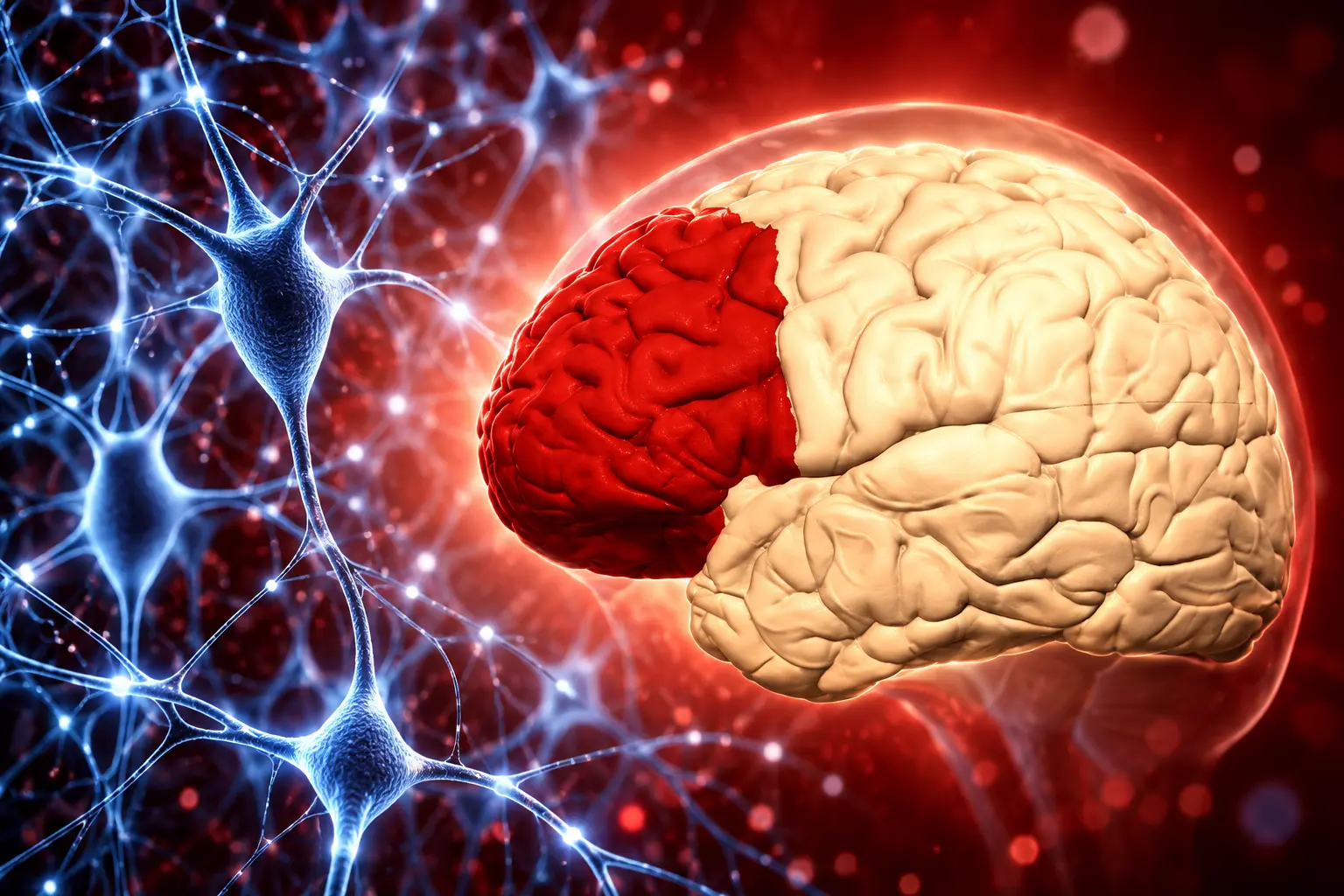
Gene Activity Settles at 24, Then Moves Again at 60
The aging paper, a lifespan single-cell transcriptomic atlas led by Hui Yang and colleagues, used more than 1.3 million nuclei from 284 people without the target diagnoses, aged 0 to 97. The team picked age 24 as the breakpoint after which the mix of most neuronal and glial subclasses stays largely steady.
That midlife plateau is long. From 24 to about 60, a span of 36 years, the cortex looks comparatively quiet at the level of gene activity. Juan Lerma, a CSIC research professor at the Instituto de Neurociencias de Alicante, said the 24-year mark confirms that cortical maturation runs past adolescence and into the mid-20s.
THREE PHASES IN THE DORSOLATERAL PREFRONTAL CORTEX
| Phase | Ages in the 284-donor set | What the cells do |
|---|---|---|
| Early remodeling | 0 to 24 (neonatal n=9, childhood n=11, adolescence n=33, plus young adults) | Neurogenesis, gliogenesis, synapse remodeling, and programmed cell death run hot. |
| Midlife stability | 24 through the 50s (young n=54, middle n=95) | Subclass mix levels off; neurons keep coordinated 24-hour clock-gene rhythms. |
| Late reactivation | about 60 and older (late adulthood n=82) | Astrocytes, oligodendrocytes, and microglia raise stress, housekeeping, and immune programs; neuronal clocks fade. |
Kiran Girdhar, an assistant professor at Mount Sinai and co-senior author of the lifespan paper, described what happens to daily timing after 60.
In young and middle-aged adults, neurons exhibit tightly coordinated 24-hour rhythms governed by core circadian clock genes. After age 60, those neuronal rhythms largely disappear, while the brain’s immune cells acquire new rhythmic activity associated with cellular stress and inflammation.
Kiran Girdhar, assistant professor of psychiatry and genetics, Icahn School of Medicine at Mount Sinai
Those clock patterns were rebuilt from times of death across about 200 neurotypical adults, not from sleep logs in living people. Roussos said they generate hypotheses about circadian aging rather than measure anyone’s night. Marta Nieto, who leads the Cerebral Cortex Development group at Spain’s National Center for Biotechnology, said the late shift moves the aging question off neurons and onto the cells that feed and guard them.
Disease genes do not sit evenly on that calendar. Genes tied to schizophrenia and bipolar disorder are most active in early development, years before typical diagnosis. Alzheimer’s-linked genes show up later in aging glia. The atlas is a clock for vulnerability, not a single portrait of “the adult brain.”
Four Dementias Line Up on the Same Cellular Faults
Donghoon Lee, assistant professor at Mount Sinai and first author of the flagship disease paper on transcriptomic vulnerability across brain disorders, said many conditions share molecular pathways and still keep distinct cellular signatures. The shared stretch is strongest among Alzheimer’s disease, diffuse Lewy body disease, vascular dementia, and Parkinson’s disease, in programs for nerve-cell development, neuronal communication, and blood-vessel biology.
Roussos had a practical reason for pooling diagnoses. Studying disorders one by one can hide biology they share, he said, while stitching together datasets built with different methods can create fake disease effects. PsychAD sequenced one region, one way, across eight named conditions: Alzheimer’s disease, dementia with Lewy bodies, Parkinson’s disease, vascular dementia, tauopathy, frontotemporal dementia, schizophrenia, and bipolar disorder.
LARGEST DIAGNOSIS GROUPS IN THE 1,494-DONOR COHORT
| Diagnosis group | Donors (paper count) | Cellular pattern reported |
|---|---|---|
| Alzheimer’s disease | 519 | Immune and vascular cells track worse cognition; interneurons associate with milder courses. |
| Schizophrenia | 177 | Deep-layer excitatory neurons are more abundant in psychiatric cases as a class. |
| Diffuse Lewy body disease | 112 | Aligns with Alzheimer’s, vascular dementia, and Parkinson’s on synaptic and vessel programs. |
| Vascular dementia | 85 | Shares the dementia cluster above, including blood-vessel gene activity. |
| Bipolar disorder | 72 | Grouped with schizophrenia on the neuropsychiatric side of the split. |
| Parkinson’s disease | 48 | Joins the four-dementia overlap on neuronal communication programs. |
Those counts overlap. Many donors carried more than one diagnosis, so the rows are not exclusive people. Non-neuronal cells are more abundant in neurodegenerative cases. Deep-layer excitatory neurons are more abundant in psychiatric cases. After the shared signatures are set aside, the remaining Alzheimer’s signal still splits by phenotype, including a link between neuropsychiatric symptoms and deep-layer excitatory neurons.
Lee’s group also traced Alzheimer’s along a progression path rather than a yes-or-no label. Early and late stages recruit different cell types. A companion Nature Medicine tool, PASCode, scores cell states against plaques, cognitive decline, resilience, and symptoms such as depression. Another paper, iBrainMap, builds donor-level communication graphs, a reply to the fact that two people with the same clinical label can look unalike in tissue.
Where Inherited Risk Shows Up in Single Cell Types
Large DNA studies have piled up risk variants for these illnesses without saying which cell should be the drug target. Georgios Voloudakis, associate professor at Mount Sinai and co-senior author of the transcriptome-wide association paper, said bulk brain averages hide those effects. The PsychAD genetics papers put the variants back onto cell types.
Biao Zeng’s Nature Genetics atlas used 5.6 million nuclei from 1,384 genotyped donors, 493 of them (35.6%) of non-European ancestry. It found 10,913 genes whose activity tracks nearby DNA variants in excitatory neurons, and only 414 such genes in endothelial cells. For Alzheimer’s, 16 genes colocalize with risk in immune cells, driven largely by microglia, with 9 in oligodendrocytes, 12 in astrocytes, and 6 in neurons, and little overlap among those lists. Microglia are not the whole genetic story.
Sanan Venkatesh and colleagues then built 94 cell-type-specific models that impute genetically regulated expression and tested them against 12 brain-disorder genome-wide association studies, with follow-up in the Million Veteran Program. They report thousands of gene-trait links that bulk tissue missed, resolved to neuronal, glial, and immune populations. Zeng’s map also ties more than 14,000 genes to cell-type-specific regulation.
Roussos put the clinical bet in plain language. A useful treatment, he said, needs to hit the right process in the right cells, and the map narrows that search by flagging vulnerable populations and processes tied to preserved function. That is a targeting claim, not a trial result.
Mount Sinai, Rush, and NIMH Supplied the Brains
The atlas exists because three autopsy programs handed over dorsolateral prefrontal cortex. The Mount Sinai NIH Neurobiobank supplied 1,042 donors, the NIMH Intramural Human Brain Collection Core 300, and the Rush Alzheimer’s Disease Center 152. Less than 5% of the Mount Sinai or Rush samples had been through single-nucleus RNA sequencing before.
WHERE THE 1,494 BRAINS CAME FROM
- Mount Sinai bank: 1,042 donors, the core of the New York collection.
- NIMH HBCC: 300 donors, including many with psychiatric diagnoses.
- Rush Alzheimer’s Disease Center: 152 donors from long-running aging cohorts in Chicago.
Donghoon Lee, posting as the flagship’s lead author, thanked the donors and families when the issue landed with a Nature cover. Eric Nestler, dean of the Icahn School of Medicine at Mount Sinai, pointed to the same collaboration across thousands of brains. Gabriel Hoffman’s group released two analysis tools, dreamlet for differential expression and crumblr for cell-composition tests, built because millions of cells from thousands of people break older statistics.
The PsychAD single-nucleus RNA-seq dataset is posted through the AD Knowledge Portal, with raw FASTQ files and processed count matrices for all 1,494 donors, plus clinical metadata. CELLxGENE hosts a browser view. Some downloads need access approval.
Ancestry is broader than older European-only brain RNA studies, and Seth Ament, a professor of psychiatry at the University of Maryland School of Medicine who reviewed a paper, said this is the first time this style of analysis has been run at this scale in people of non-European descent. It is still a majority-European sample. A Cell Genomics audit of several single-cell resources, including PsychAD, found Asian representation under 2% in that consortium’s slice. Scale and representation are not the same job.
Frozen Nuclei Cannot Explain a Living Illness
Every nucleus in the map is from a dead brain. Ament said what is present in that tissue reflects a whole life, and for complex disease the main use is to generate hypotheses about where to look next. Nieto made the same cut: the atlas shows which programs change with age, not whether glial stress causes neuronal failure, follows it, or protects against it.
The map is also one region. Roussos said additional areas will be needed for a full picture. RNA at death does not capture proteins, connections, or a living 24-hour cycle in the same person. Psychiatric diagnoses in autopsy series are messy, and comorbidity is common, which is why the team modeled overlap instead of pretending each brain had one label.
WHAT WE KNOW
- The calendar: prefrontal gene activity has a developmental burst, a long plateau from age 24, and a glial-heavy shift at about 60.
- The overlap: four dementias share synaptic and vascular programs in this region; psychiatric cases skew toward deep-layer excitatory neurons.
- The genetics: inherited risk is cell-type specific, and bulk tissue hid a large share of the links.
WHAT IS UNCONFIRMED
- Causality: postmortem RNA cannot prove that late-life glial programs drive dementia.
- Other regions: hippocampus, temporal cortex, and subcortex are not in this map.
- Treatments: no trial has used these cell lists as drug targets yet.
Online recaps of the papers keep making that last point in different words: this is a high-resolution street map of donated tissue, not a test and not a medicine. Roussos said the next steps are more regions, more ancestries, other molecular layers, and experiments that test the mechanisms the atlas only ranks. For now the public resource is the product, and the timetable it gives, from a noisy first act through a long quiet and a second remodeling at about 60, is what the 1,494 brains actually changed.
Frequently Asked Questions
Where can researchers access the PsychAD dataset?
Raw FASTQ files and joint h5ad count matrices for all 1,494 donors sit in the AD Knowledge Portal study folder, with clinical and assay metadata, and CELLxGENE offers an interactive taxonomy view; some files require a data-use application before download.
What is single-nucleus RNA sequencing in this study?
The team isolated nuclei from frozen dorsolateral prefrontal cortex because intact cells rarely survive freezing, then sequenced RNA still in each nucleus, which records gene activity at death but misses most cytoplasmic transcripts and any living-cell dynamics.
Which grants funded the PsychAD Consortium?
The lead National Institute on Aging award is R01AG067025 to Panos Roussos and Vahram Haroutunian, with additional NIH awards including R01AG082185, R01AG078657, R01AG065582, R01MH125246, and U24AG087563 listed on the genetics papers.
What does PASCode add for Alzheimer’s research?
PASCode is an artificial-intelligence scorer trained on more than 6 million nuclei that links individual cell states to Alzheimer’s pathology, cognitive decline, resilience, and neuropsychiatric symptoms, including features that overlap with depression rather than with plaques alone.
How many genes show DNA-linked activity in excitatory neurons?
The Nature Genetics eQTL atlas reports 10,913 such genes in excitatory neurons at the class level, against 414 in endothelial cells, and 8,812 in layer 2/3 intratelencephalic excitatory neurons at the finer subclass level.
Disclaimer: This article is news reporting on published research and is for information only. It is not medical advice, a diagnosis, or a recommendation to start, stop, or change any treatment for Alzheimer’s disease, Parkinson’s disease, schizophrenia, bipolar disorder, or any other condition. Readers who have symptoms or treatment questions should speak with a qualified physician or neurologist before acting on any finding described here. Cell counts, donor figures, and disease labels reflect the PsychAD papers and consortium materials as released on 23 September 2026 and may be updated as the public dataset is revised.

OpenAI’s Agent Monitor Waited During the Hugging Face Breach

Brent Hits $116 as Recovered Gulf Oil Draws Attacks

Hellebuyck Lands in New Jersey on a Five-Year Cup Bet

Xbox Plans Three More Layoff Rounds Before Helix

OpenAI’s $30 Billion Round Ties UAE Cash to Gulf Compute

Consumer Sentiment Falls to 51.7 as Future Outlook Darkens

Pluto’s Heart Glacier Still Pushes Liquid Nitrogen Upward

The Extra Heat Month Hits Children Where Care Is Thinnest

Take-Two Subpoenas Microsoft While GTA 6 Preorders Soar

Abbott Pays $670 Million to Exit a Missouri Food Verdict
-
 BUSINESS2 months ago
Consumer Sentiment Falls to 51.7 as Future Outlook Darkens
-
 NEWS2 months ago
Pluto’s Heart Glacier Still Pushes Liquid Nitrogen Upward
-
 NEWS2 months ago
The Extra Heat Month Hits Children Where Care Is Thinnest
-
 GAMING2 months ago
Take-Two Subpoenas Microsoft While GTA 6 Preorders Soar
-
 BUSINESS2 months ago
Abbott Pays $670 Million to Exit a Missouri Food Verdict
-

 NEWS2 months ago
UMMC Will Rebuild 30-Year-Old Cancer Labs With $2.4 Million
-

 ENTERTAINMENT2 months ago
Netflix Weighs Hosting Peacock and Fox One in Its App
-

 GAMING2 months ago
GTA 6’s 80-Hour Run Counts Goals That Change the Story